Assignment #3

Your name:
Your email address:

Exploring intein structures in chimera

Objectives:


Background information:

Inteins are molecular parasites that have their own life cycle. Once they are in gene in one member of the population, they spread by super-Mendelian inheritance. If as a consequence of sex of gene transfer, an infected and a non infected allele come together in one cell, the homing endonuclease activity of the intein makes a double-strand cut in the DNA of the non infected allele. During the repair of the break the intein is copied into the allele. The other activity of the intein is a self splicing activity: The DNA encoding the intein is translated and transcribed together with the host gene. At the protein level, the intein removes itself from the host protein (aka as extein).

Once an intein is fixed in a population, there is nothing left to do for the homing endonuclease. The endonuclease activity decays and may ultimately be deleted, leading to mini-inteins that only contain the self-splicing domain. For more discussion see here.

Most inteins are composed of two domains: one is responsible for protein splicing, and the other has endonuclease activity. A few inteins have lost the endonuclease domain completely and retain only the self-splicing domain and activity. The latter inteins are called mini-inteins .

The structures of several inteins are crystallized.

To do:

  1. Open 1VDE in chimera. This structure has two chains. Select chainA and save the selected residues into there own pdb file (file > save pdb > fill out the form, check save selected residues only, save). Close the session.

  2. Reopen chain A in chimera. Open Mycobacterium mini intein 1AM2. Depict the structures as ribbons and color them according to the secondary structure. Rotate the two structures until you can see the similarities between mini intein and the large intein. (use the model panel to rotate one or the other structure). Which part of the structures appears to be similar?
    Your answer --->

    Align two structures using matchmaker. Does the alignment correspond to your expectation?
    Your answer --->

    Can you find which part of Saccharomyces cerevisiae intein corresponds to the endonuclease domain by comparison of the two structures? Using tools > compare structure > match -align create a pairwise alignment of the two inteins. Color the putative self-splicing (i.e. the part that is present in 1VDE and in 1AM2) and endonuclease domains of 1VDE (no corresponding part in 1AM2) in two different colors (selecting consecutive residues works easily via the alignment window). Same result as above?
    Your answer --->

    Find and select the N and C terminals (First a.a. and the last a.a.) in both structures. If you hover over the beginning or end, the name of the residues pops up in a little window. CTRL click selects the amino acid or atom, and shift control click adds to the selection. Under actions>atoms/bonds>show side-chains make the side chains of the first and last amino acid visible. Optional: hide the rest of the structure, if it distracts you. Rotate the structure of the sidechains of the first and last aa and decide which atoms are closest. Select these atoms (ctrl click and shift ctrl click), then go to tools>Structure analyses> distances and click create. Repeat this for a few atoms from the first and the last aa. How close are beginning and end (in Ångström and in nanometers)? .
    Your answer --->
    Save your project.

  3. Open Saccharomyces cerevisiae intein that is bound to its target DNA sequence. Does the DNA - Protein interaction in 1LWS agree with your previous assignment of the self-splicing domain? (see the saved structure from the previous exercise)
    Your answer --->

  4. Try to find a way to display the interactions between the amino acid side chains and the DNA helix. One way to do it is to select two DNA chains and select aa in the neighboring zone. To do this you could first select chain A, then invert the selection (you now should have selected Chain B and C). Then select zone 4 or 5 Angstrom. Make the side chains visible, and display either the side chains or the DNA as spheres. One way to look at individual interactions is to turn the molecule so that one looks down the DNA helix, and use the viewing controls only look at a cross section (or Slab) of the structure. If the bases of the DNA are displayed too cartoonish, you can change the display in Actions > Atoms/Bonds > nucleotide objects (select different option and click apply).
    Most of the interactions of aa side chains are with the major groove of the DNA. Do you find residues that interact with the minor groove? If yes, which aa are involved:
    Your answer --->


  5. The Lys 340 and Glu 366 are residues that are important for interaction with DNA. Select those residues (Tools> sequence>sequence allows to show the primary sequence, which is a good way to select a particular aa). Which base pairs interact with these amino acids? (if you hover over an atom, a pop-up window gives the base, the number of the base, and the chain (e.g., G 23.B is the 23 rd base in chain B, which is a Guanin).
    Your answer --->

image


Comparing divergent proteins with similar structures

A) GRASP ATP binding domain

Glutathione synthetase and D-Alanine D-Alanine Ligase were long ago (1990) recognized by Jim Knox (MCB faculty) to be so similar in structure that there could be no doubt about their common evolutionary origin. Later these and other enzymes were discovered to have a novel ATP binging site (the GRASP domain). These domains were identified using profile alignments (will be covered later in this course). A description of the GRASP domain family is at https://www.ncbi.nlm.nih.gov/pmc/articles/PMC3249071/.

Save the following files to your computer and load them into chimera.

2DLN.pdb (D-Alanine D-Alanine Ligase - D-ala is an important part of the bacterial cell wall, more here, Flash animation here) [you may get a warning/error message... ignore it] ,

1GSA.pdb (glutathione synthetase from E. coli, glutathione is the biological equivalent of mercaptoethanol),

cpsBfrag.pdb, load cpsFfrag.pdb (Carbamoyl phosphate synthetase is an enzyme consisting of several domains. These are the front and the back of (1BXR)).

Based on your first impression, are these structures similar? homologous?

Your answer --->

Can you use Tools > Structure comparison > Matchmaker to align these structures?

Your answer --->

See here for an illustration of the structures in similar orientation.


Finished? Leaving the lab? Remember to...

Choose one of the following options, then click the button.

Send email to your instructor (and yourself) upon submit
Send email to yourself only upon submit (as a backup)
Show summary upon submit but do not send email to anyone.


Optional exercises:

Histones

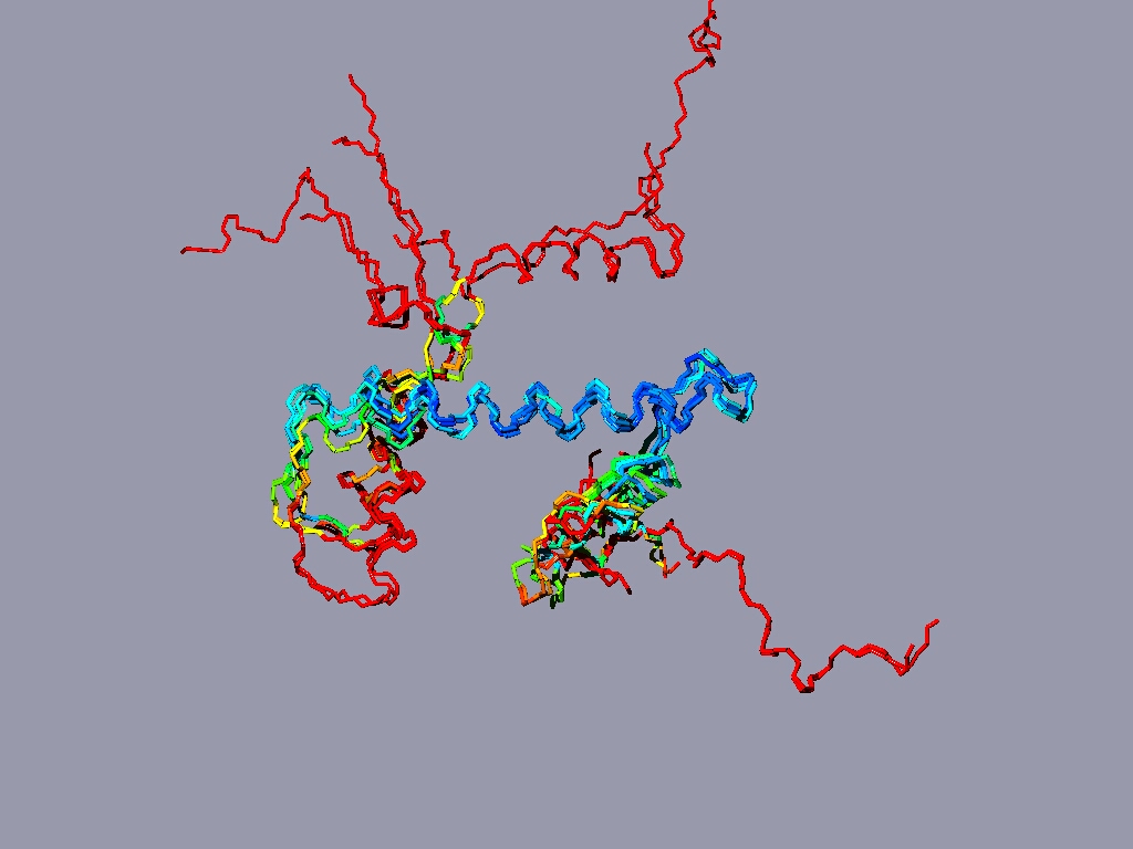
If you have more time to spare and you are up for a challenge, take a look at the nucleosome (1AOI.pdb). Open it from within chimera. Save each of the 8 histones as a separate pdb file. Close the nucleosome file, open the 8 histone files and align them to one reference histone.


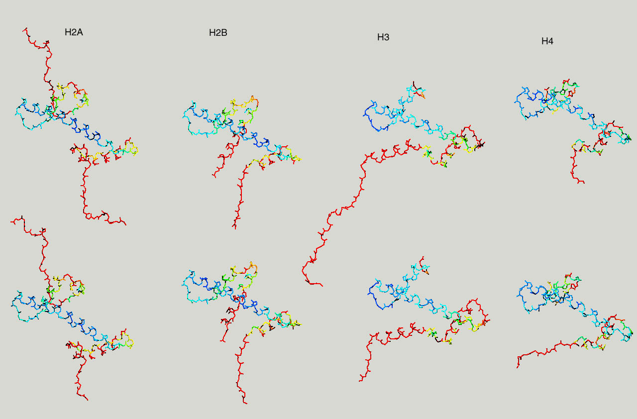
Below same as last figure, but histones are depicted side by side :


The proteasome core

Proteasomes are the recycling cans of the cell. Ubiquitin tagged protein are taken-up into the proteasome and broken down into aa. The proteasome (20S) core consists out of homologous alpha and beta subunits, arranged in 4 rings. The two central ones are beta subunits (2x7), the two rings at the ends are made from alpha subunits. Humans (and other eukaryotes) have 7 different genes encoding seven different alpha subunits, and 7 different genes encoding beta subunits, and each is present in duplicate in the 20S proteasome core. Altogether 28 subunits.

We are interested in creating a structure based sequence alignment of the different alpha and beta subunits. To do so, pick the bottom or top half of the proteasome (5LF7.pdb, or a similar high resolution structure from another organism), and save all the different subunits as separate pdb files (if you use 5LF7.pdb, the pdbs for the individual SU are here). First align all the alpha beta subunits separately, and then try to generate an alignment of the alpha with the beta subunits. Use matchmaker to align the structures, then use Tools > structure comarison > Match-Align to create a multiple sequence alignment.

proteasomeSU

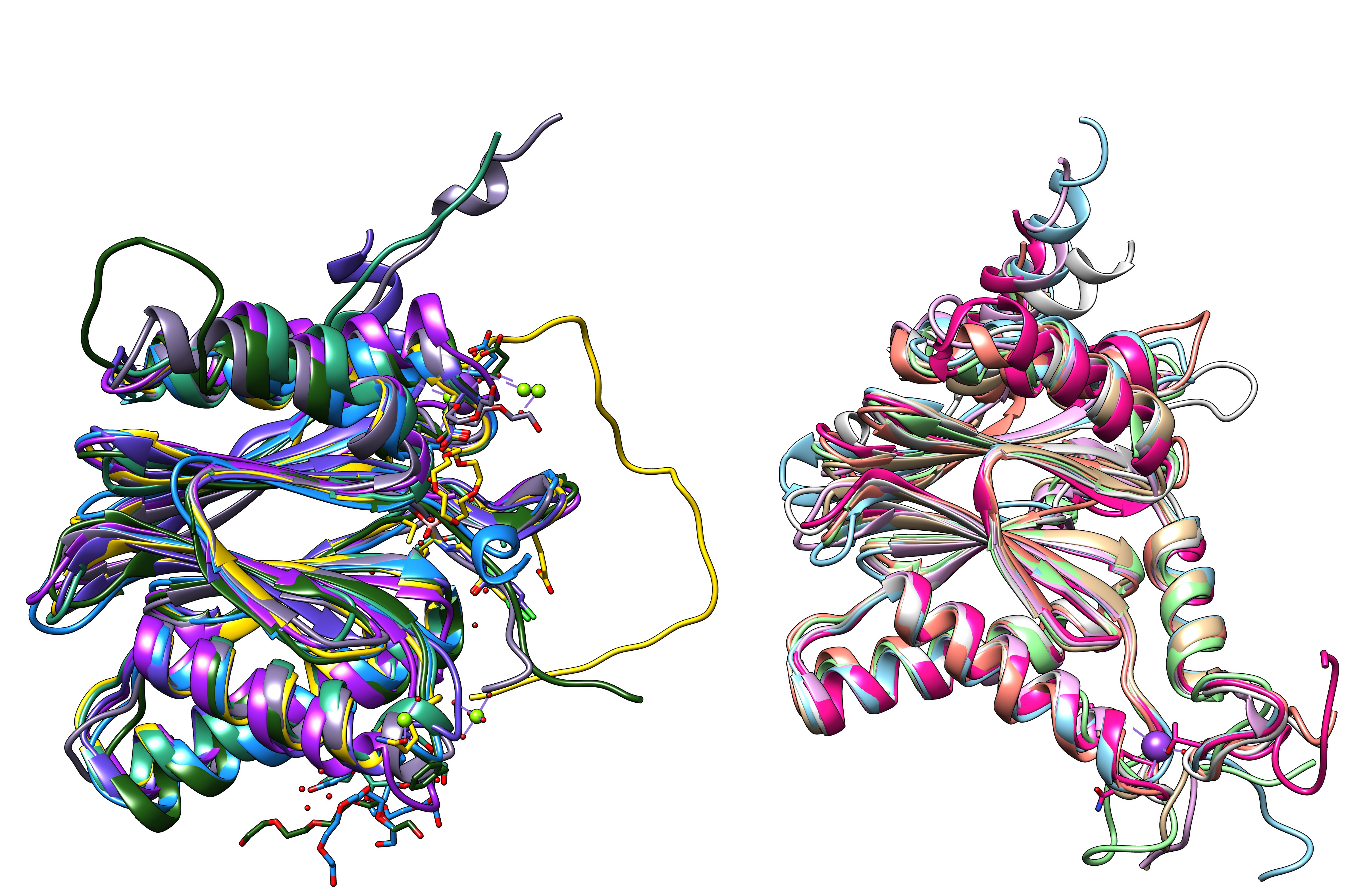
Proteasome subunits (7 beta and 7 alpha subunits) from human hela cells (5LF7.pdb)

all proteasome SUs

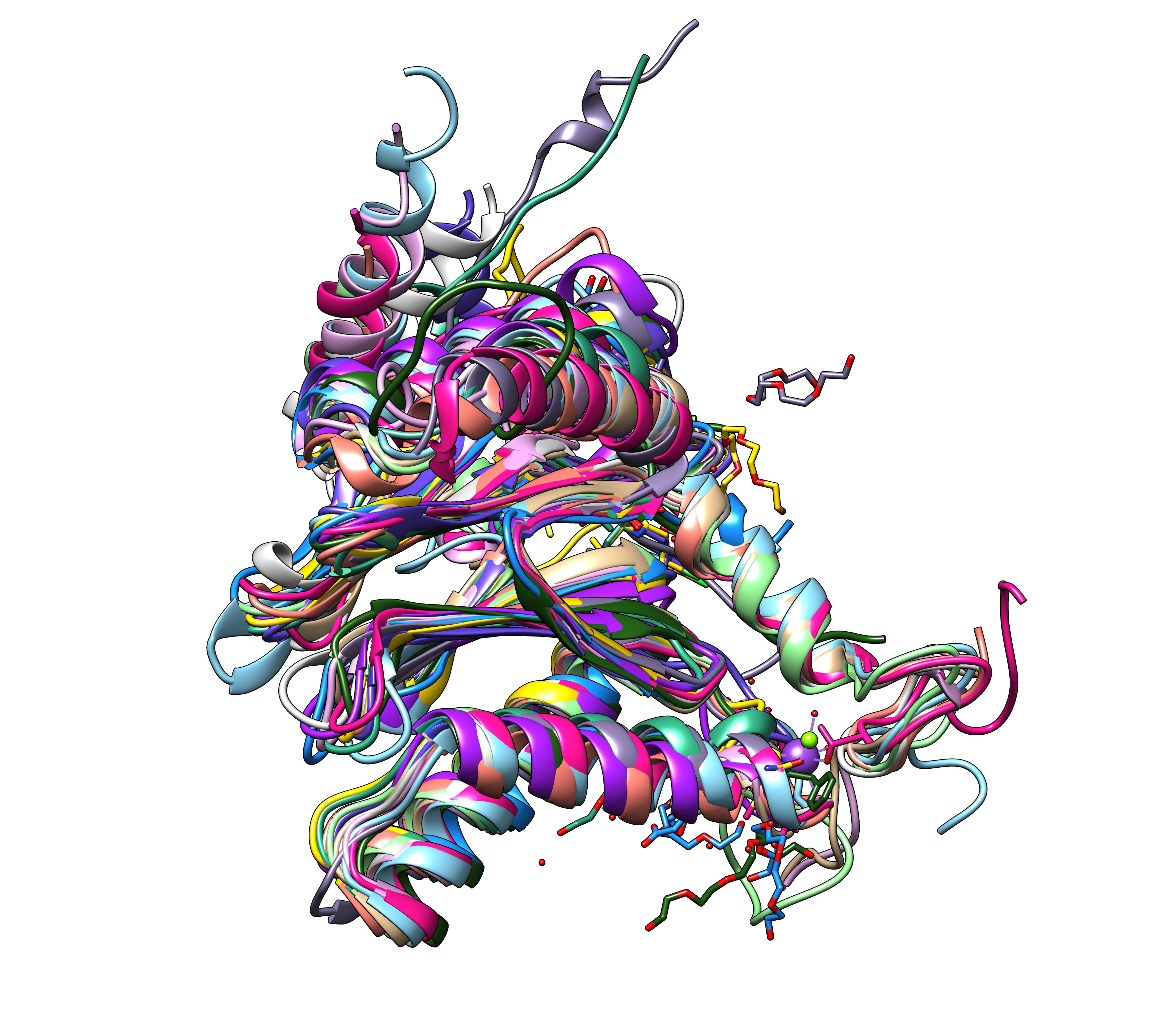
All 14 subunits of the human proteasome core superimposed.